Anatomic Pathology
Clinical Pathology
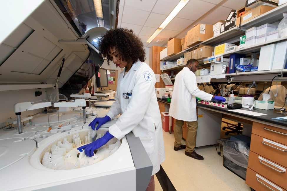
Clinical pathology services include: hematology (complete blood counts with white blood cell differentials, serum biochemical panels, hormone levels); parasitology; microbiology (bacterial and fungal identification, antimicrobial sensitivity); analysis of urine and other body fluids; serology for numerous bacterial, viral and parasitic infectious agents; and, environmental monitoring. All tests are performed by qualified laboratory professionals trained in veterinary laboratory techniques. The laboratory staff is committed to research in improved diagnostics, reference range development and state-of-the art assays. New tests are continuously being added as experimental needs change. The LCP also utilizes a network of reference laboratories to provide tests that are not performed in-house.
Histotechnology
Subcutaneous hemangiosarcomas in a P53 homozygous knockout mouse. Hematoxylin and Eosin, 40x. Courtesy of Dr. Krista La Perle.
Immunohistochemistry
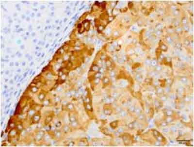
Chromogranin A, mouse adrenal, pheochromocytoma
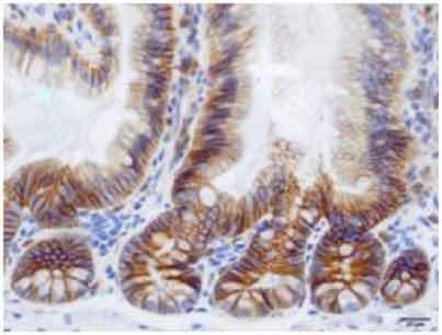
E-cadherin, mouse small intestine, normal
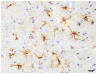
GFAP, mouse brain, normal
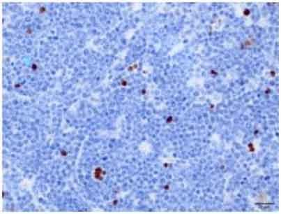
Cleaved caspase-3, mouse lymph node, lymphoma
Cell type markers
|
Proliferation markers and cell cycle
Apoptosis and anti-apoptosis markers
Angiogenesis markers
Tags and reporters
Others
|
m: mouse specific, h: human specific
- Unless indicate, stains are optimized and validated for formalin or paraformaldehyde fixed paraffin embedded (FFPE) mouse tissues. For staining of samples prepared differently, or from other species, please consult the laboratory before submitting samples.
- For all immunofluorescence, please consult the laboratory before submitting samples.
- For advice on sample preparation and selection of appropriate markers, please consult an LCP technician or pathologist prior to submission.
- Evaluation of IHC slides by an LCP pathologist is available; additional charges apply for this service.