Now Accepting Samples

MALDI Imaging

Overview

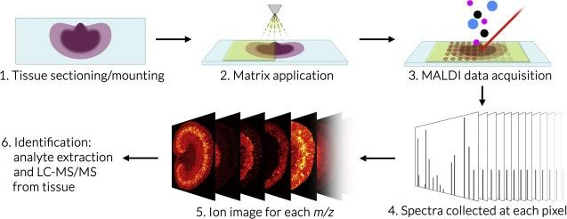
Imaging mass spectrometry (IMS) is a sensitive molecular technique that uses mass spectrometric detection of biomolecules to generate quantitative spatial distribution maps that can be overlaid with optical images. IMS enables the simultaneous analysis of thousands of different molecules such as metabolites, lipids, and peptides in a single experiment without labeling. 

Imaging Mass Spectrometry Schematic Overview

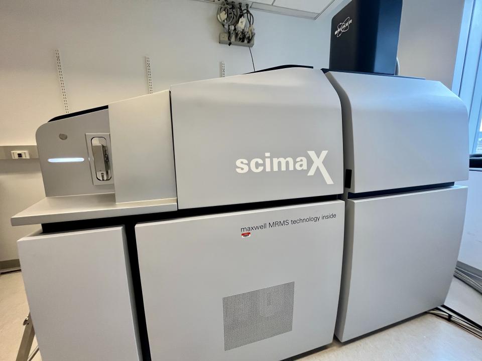
The Bruker SciMax is an ultra-high mass resolution Fourier Transform – Ion Cyclotron Resonance (FT-ICR) mass spectrometer capable of determining fine isotopic structure that enables unambiguous identification of biomolecules. The SciMax uses laser ionization (MALDI) to raster across a tissue slice collecting mass spectra pixel by pixel. Each spectrum contains information that can identify and provide relative quantification for hundreds or even thousands of different molecules. By adjusting the size and spacing of laser pulses, the SciMax can generate images with differing spatial resolutions. We offer high (25 µm) and low (80 µm) resolution.

MALDI Imaging Services are billed by hours of instrument and subsequent analysis time with a modest fee for slide coating with matrix. For high spatial resolution analysis, it is important to identify areas of interest.
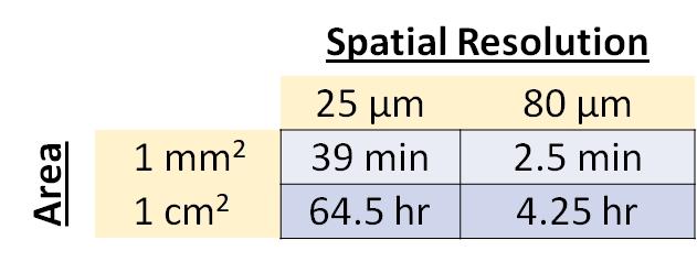
IMS Time Per Area

***All IMS Services require prior consultation with the core.

For inquiries, please contact:

Dr. Shu Zhang (shz4010 at med.cornell.edu).

Sample Preparation

High quality sectioning and mounting is essential for success. However, the core is currently unable to provide tissue sectioning and mounting services. Users must prepare samples on their own or through a histology core facility following the procedure detailed in the Sectioning and Mounting Guide (link to pdf below). 

General Considerations

Samples must be mounted on special conductive slides coated with Indium Tin Oxide (ITO). We recommend CB-90IN-S111 (25 mm x 75 mm x 1.1 mm) from Delta Technologies.

To prepare cryo-sections (8-14um), it is best to mount sections that you wish to compare (e.g. different genotype or treatment with replicates) on the same  slide. There is no particular limit on the number of sections per slide (we have seen 12 brain sections on one slide). However, you MUST leave a margin of ~5 mm on the long ends of each slide.

Slide Margins

Always prepare extra slides in case experiments fail.  For survey experiments, we need at least 2 slides - one for positive and one for negative ion mode. Prepared slides should be stored at -80°C or under vacuum at RT (up to 1 month) until MALDI MS experiment.

Analyzed slides can be washed for subsequent H&E staining and optical imaging. Optical images can then be co-registered with MALDI images. We are not currently equipped to perform the H&E staining. Users must take the slides back or prepare adjacent serial sections for H&E.