Citigroup Biomedical Imaging Center (CBIC)

Summary 

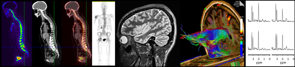
The WCM CLC Citigroup Biomedical Imaging Center (CBIC) provides state-of-the-art imaging instruments and expertise in their applications to the Weill Cornell Medicine (WCM) community and to outside investigators.  Resources and services include magnetic resonance imaging (MRI), positron emission tomography (PET), single photon emission computed tomography (SPECT), X-ray computed tomography (CT), high resolution ultrasound, and optical imaging and endoscopy.  The CBIC provides consultation on project design and image visualization and analysis, and offers seminars, training and educational workshops.


Services

Magnetic Resonance Imaging (MRI): MRI is used to study a wide range of diseases in patients, from neurological and psychiatric disorders to cancer and vascular disease. Additionally, MRI imaging is performed on both large and small live animals to investigate disease-related functional and structural changes.  Applications include angiography, diffusion tensor, functional, perfusion and spectroscopic imaging.  MRI supports high resolution imaging virtually anywhere in the body.  Instruments in the CBIC include: Siemens 3.0 Tesla PRISMA MRI with 64 high bandwidth receivers providing fast parallel image acquisition with a 80 mT/m gradient amplitude; GE Discovery 750 MRI with 32 high bandwidth receivers and a 50 mT/m gradient amplitude (oth scanners have a 200 T/m/s slew rate and a 60 cm inner bore diameter); Bruker BioSpec 70/30 USR 7.0 Tesla Small Animal MRI with a 200 mT/m gradient amplitude, a 640 mT/m/s slew rate and a 20 cm inner bore diameter (in-house built RF coils provide additional signal and localization compared with standard imaging coils).  

Positron Emission Tomography/Computed Tomography  (PET/CT): Instruments in the CBIC combine sophisticated computed tomography (CT) and positron emission tomography (PET) to produce images that simultaneously provide anatomic and metabolic information.  MicroPET is available for small animal imaging studies.  PET can be used to scan the entire body or selected organs.  PET precisely measures physiologic function, detects metabolic changes in tissue, displays blood flow, and tracks alterations in biochemical processes.  PET can help physicians evaluate patients for coronary artery bypass or angioplasty procedures, diagnose psychiatric and neurological diseases, assess head trauma and movement disorders, and help diagnose and stage tumor malignancies.

Cyclotron / Radiochemistry: A medical cyclotron (Ebco 19 MeV Dual Beam Cyclotron) is available for production of radiotracers and radiochemistry equipment is available for ligand synthesis. The cyclotron produces various positron-emitting radiolabeled pharmaceutical drugs that are designed and engineered to complement clinical molecular targets.  The preparation of these unique drugs requires a source of radionuclide (such as 11C or 18F) and the tools for subsequent synthesis of the drug incorporating the radionuclide. The cyclotron produces 19.2 MeV protons and 9.5 MeV deuterons in two separate beam lines. 

Optical Imaging: The Bruker In-Vivo XTREME is a powerful and versatile small animal imaging system for preclinical research that includes multispectral fluorescence, luminescence, radioisotopic and high resolution X-ray imaging in one system.  The IVIS SpectrumCT enables simultaneous molecular and anatomical longitudinal studies, providing researchers with essential insights into complex biological systems in small animal models. 

High Resolution Ultrasound:  The VisualSonics Vevo 3100 is located in a vivarium and provides noninvasive in vivo measurement of anatomical structures, cardiac motion and blood flow.  Features include transducers with 40 µm, 50 µm, and 75 µm resolution, 20 mm field of view, and image-guided needle injection.

Image Visualization and Image Analysis: Available image analysis software includes ImageJ (NIH).  Functional MRI (fMRI) analysis may be performed using AFNI or SPM.  Diffusion tensor imaging (DTI) may be processed using the Siemens Syngo workstation, DTI Studio or TrackVis.  PET pharmacokinetic analysis is performed using PMOD software with multiple toolboxes.  Advanced applications programming is performed in MATLAB, IDL and ImageJ routines.  Data processing is performed on PC, Mac, Silicon Graphics and Linux based systems.  Dedicated analysis machines include a Linux workstation with an NVIDIA Tesla K20C, 5GB, Graphics Processing Unit containing 2,496 processor cores for parallel analysis.  Online data storage is maintained on a secure 8.5 Tb partition on a WCM ITS research storage system.


Resources

CBIC major equipment includes two 3 Tesla magnetic resonance imaging and spectroscopy (MRI/MRS) systems for humans and large animals; a 7 Tesla/30cm bore pre-clinical MRI/MRS system; a combined positron emission tomography and computed tomography (PET/CT) system for humans and large animals; a pre-clinical multi-modality PET/SPECT/CT system; two ultra-high resolution CT systems; a multispectral optical imaging system including bioluminescence, fluorescence, and X-ray; a medical cyclotron facility for production of radiotracers; and radiochemistry equipment for ligand synthesis.  In addition, the CBIC supports a BSL-2 compliant imaging pre-clinical imaging facility that includes high resolution ultrasound, optical/CT, PET, and endoscopic imaging systems. 

Magnetic resonance imaging and spectroscopy (MRI/MRS): Two 3.0 Tesla systems for whole body human and large animal studies (General Electric Discovery 750 3.0 Tesla, Siemens PRISMA Fit 3.0 Tesla), Bruker BioSpec 70/30 7.0 Tesla / 30 cm bore for pre-clinical studies, and a small bore 1.0 Tesla 1.0 Tesla ASPECT M3 system for small animal studies. 

Positron emission tomography (PET), single photon emission computed tomography (SPECT), and computed tomography (CT): Pre-clinical multi-modality PET/SPECT/CT system (Siemens Inveon), pre-clinical PET (Siemens Focus 220), pre-clinical CT (Scanco Viva CT 40), high resolution specimen CT (Scanco microCT 35), whole human body ultra-high resolution PET/CT (Siemens Biograph). 

Radiochemistry: Medical cyclotron (Ebco 19 MeV Dual Beam Cyclotron) for production of radiotracers; radiochemistry equipment for ligand synthesis. 

Optical imaging: Bruker Xtreme multispectral optical imaging system (including bioluminescence, fluorescence, and X-ray), optical CT (Perkin Elmer IVIS SpectrumCT Imaging System). 

Ultrasound: VisualSonics Vevo 3100 Imaging System. 

Endoscopy: Storz endoscopy system.


More Information

Publications

Contact Us: 

Douglas Ballon, Ph.D.
Director, CLC Citigroup Biomedical Imaging Center
(212) 746-5679
djb2001@med.cornell.edu

Front Desk
CLC Citigroup Biomedical Imaging Center
(212) 746-5889

For more information about services and pricing, to register as a user of the core, to submit samples, to schedule time on the instruments, and/or to arrange for training, please go to the WCM iLab portal at http://wcmc.corefacilities.org.

For more information about the CBICs, please visit the Citigroup Biomedical Imaging Center website.