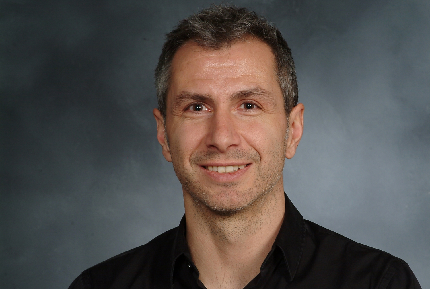
A three-year, million-dollar grant from the W.M. Keck Foundation will fund an ambitious project, led by Weill Cornell Medicine, to develop a next-generation, high-speed atomic-force microscope (AFM), capable of capturing the dynamics of proteins in unprecedented detail.
The W. M. Keck Foundation was established in 1954 in Los Angeles by William Myron Keck, founder of The Superior Oil Company. One of the nation’s largest philanthropic organizations, the W. M. Keck Foundation supports outstanding science, engineering and medical research.
“We’re very excited about this award and this project—our aim is to improve the speed of AFM ‘movies’ by two orders of magnitude over current commercial technology,” said project leader Dr. Simon Scheuring, professor of physiology and biophysics in anesthesiology at Weill Cornell Medicine. “This will allow us to record protein dynamics that cannot be seen with current technology but can be crucial to understanding how these proteins work and how to target them with drugs.”
AFM is different from most other types of microscopy in that it doesn’t illuminate molecules of interest with photons of light or electrons. Instead, it uses an exquisitely tiny, oscillating probe to “feel” the surface of the molecule and thus construct an image of it—much as a blind person constructs mental images by touch.
AFM has the key advantage that it can continuously scan the surface of a protein, in theory at high enough speed to record changes in the shape or “conformation” of the protein. Proteins often exist in different conformations as they work. For example, ion-channel proteins, which are fundamentally important in many biological processes, have multiple conformations including “open” and “closed” conformations. In many cases, one cannot really know how a protein works—and cannot optimally target it with drugs—unless one knows the full details of its conformations, its short-lived transition states and the pathway between them.
The current standard methods for imaging protein structures at atomic or near-atomic scale, X-ray crystallography and electron microscopy, have severe limitations for studying protein dynamics. These techniques calculate structures through “averaging” hundreds of thousands of molecular snapshots to build up a clear image of a protein. In this averaging process, only the most dominant and stable protein conformation is resolved, whereas more fleeting conformations are lost.
Existing commercial high-speed AFM technology is better, but so far can only resolve protein conformations that last on the order of the second. Dr. Scheuring and his laboratory, in experiments in the past few years, have been able to push this temporal resolution into the range of a hundred milliseconds. Under the new project, they expect to push this limit further, to the millisecond range, opening an enormous opportunity for new discoveries about protein dynamics.
Participants in the project will include postdoctoral researchers Drs. Atsushi Miyagi, Shifra Lansky, Alma Perrino, and Runze Ma from the Scheuring laboratory in the Weill Cornell Medicine Department of Anesthesiology, and collaborating scientists from Weill Cornell Medicine, Rockefeller University, the Perelman School of Medicine at the University of Pennsylvania, and Washington University at St. Louis.
One of the strategies for achieving this “next-generation high-speed AFM” tech includes the use of a dual measurement system for recording, respectively, more coarse and more fine details on a protein surface. In this installment, the coarse measurement system can run at increased speed over the globular biological structure, while a high-speed sensor records the delicate features. Another development concerns a circular scanning method, rather than the traditional side-to-side, raster-type method. In this mode, the coordinated action of proteins can be visualized with sub-millisecond temporal resolution.
“We plan to demonstrate this initially on ion-channel proteins, but our technology ultimately should have very broad applications for studying protein dynamics—indeed, it could revolutionize biomedical imaging,” said Dr. Scheuring who is also a professor of physiology and biophysics at Weill Cornell Medicine.
