Microscopy and Image Analysis
Core Facility


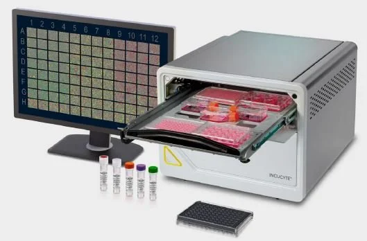
NEW EQUIPMENT COMING IN EARLY 2024
(Click images for details)
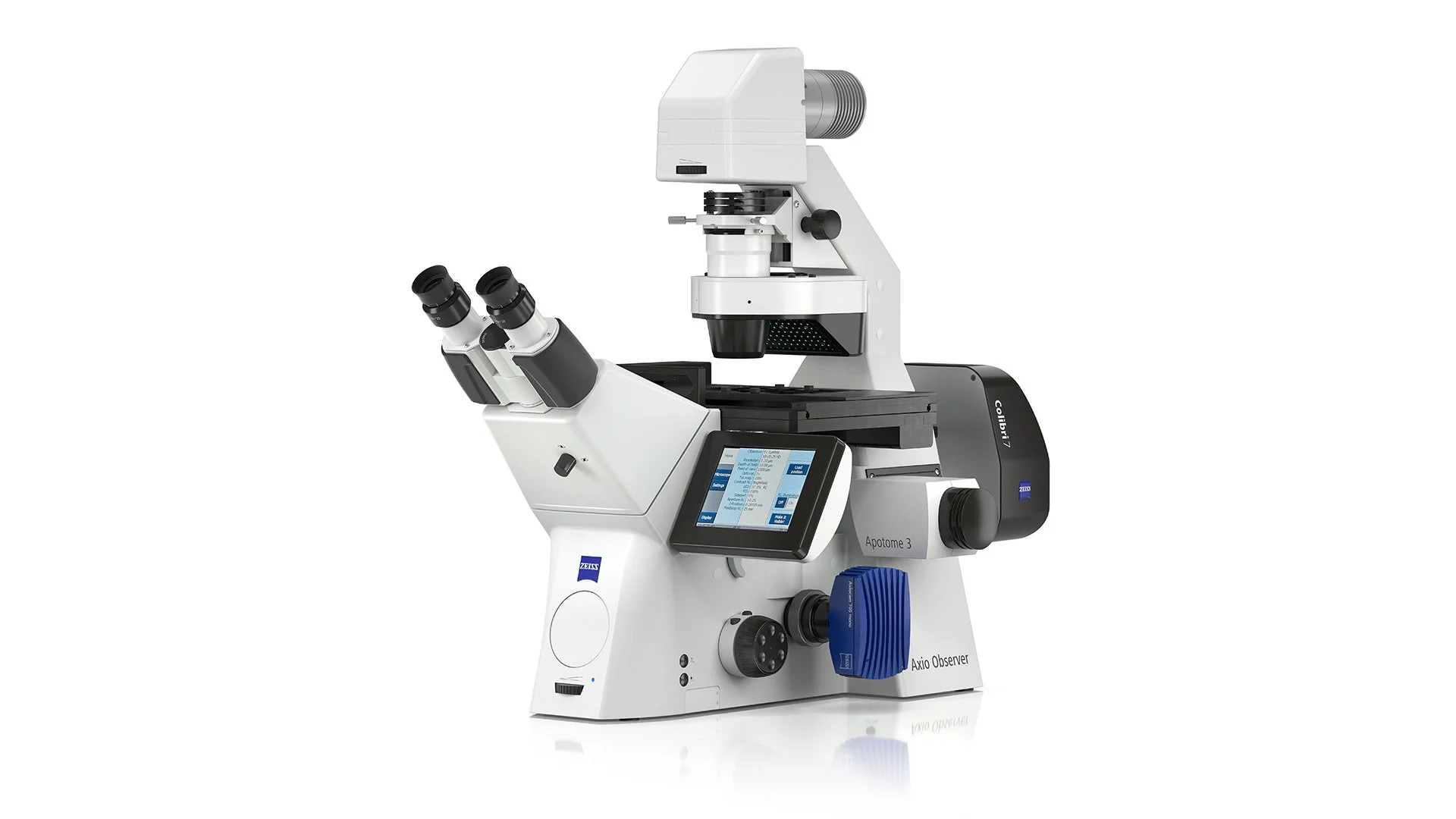
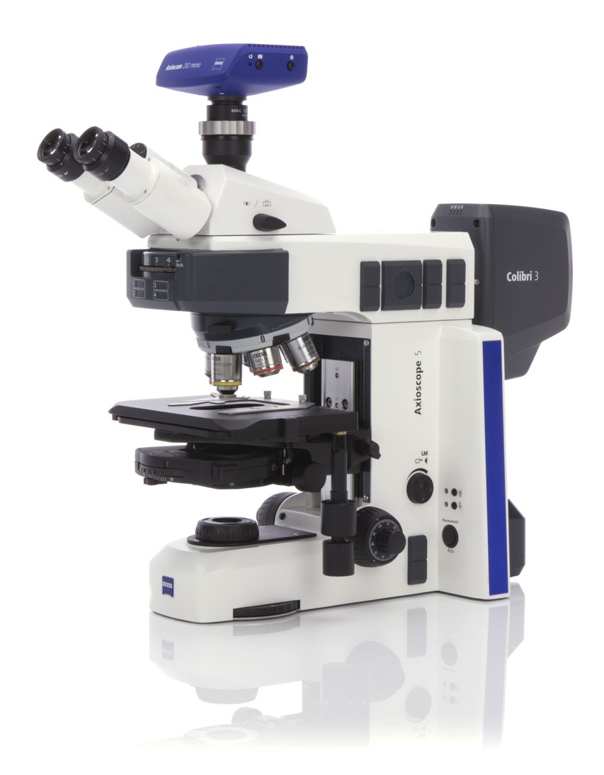
EQUIPMENT ADDED IN 2023
(Click images for details)

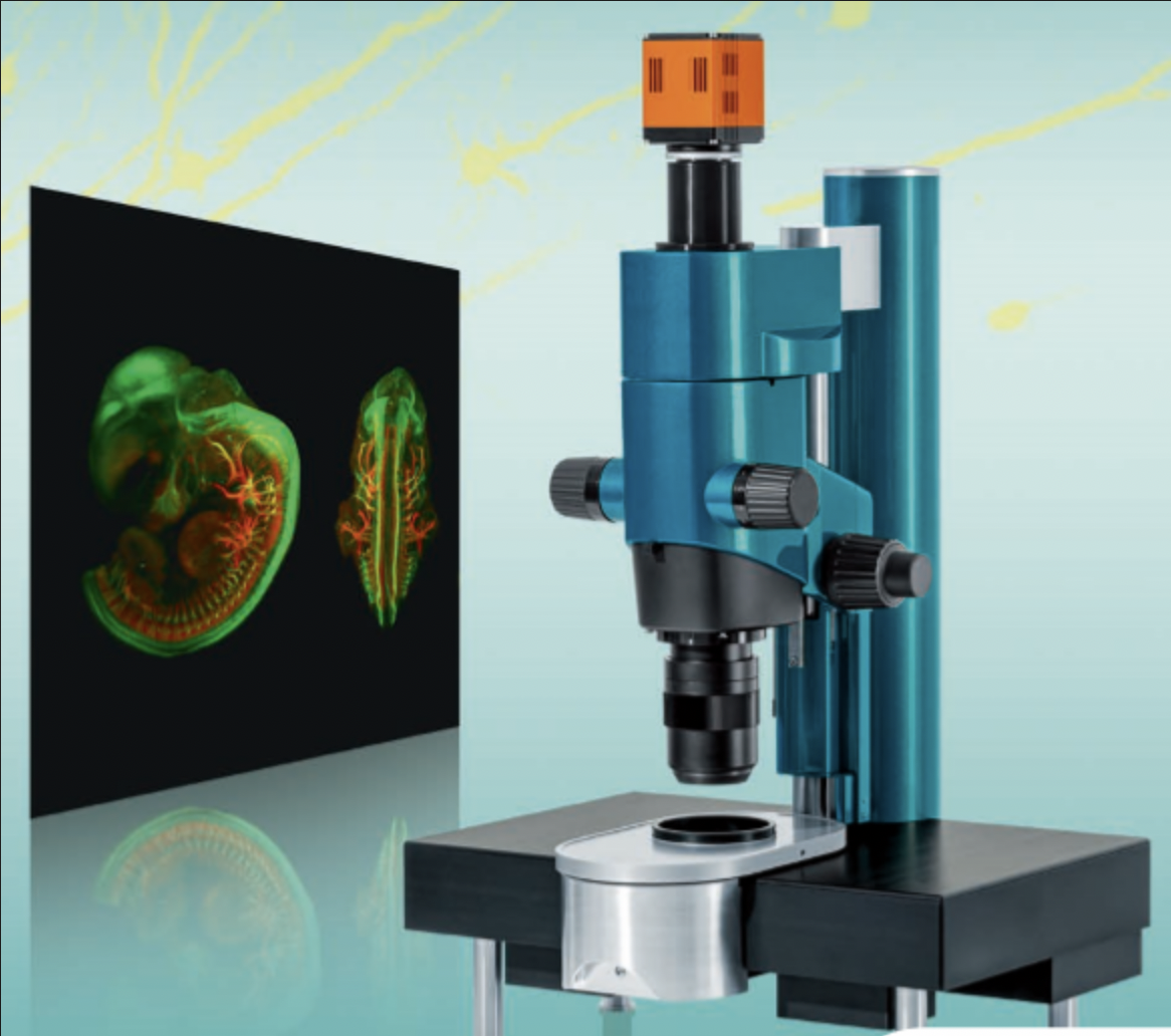
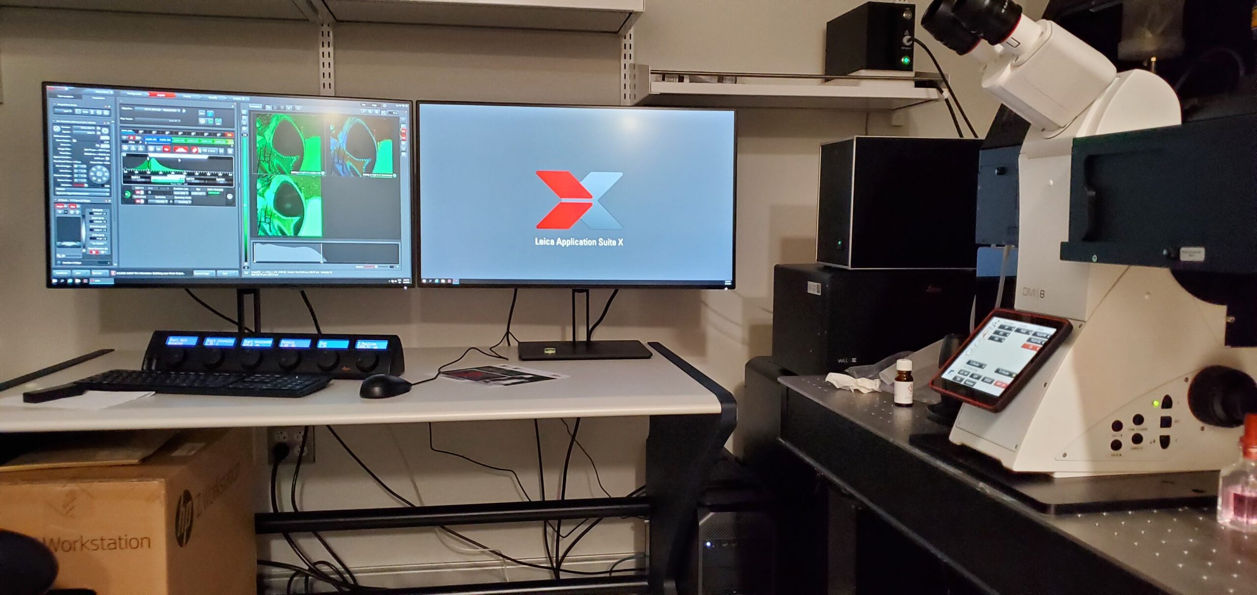
EQUIPMENT ADDED MID-LATE 2022
(Click images for details)

Additional advanced microscopy services
available through Weill Cornell Medicine’s institutional membership at the link below
The Imaging Core Facility strives to assist users with all their microscopic imaging needs, from the conception of an idea, to final image analysis and presentation. Our specific offerings are listed below.

Microscopy
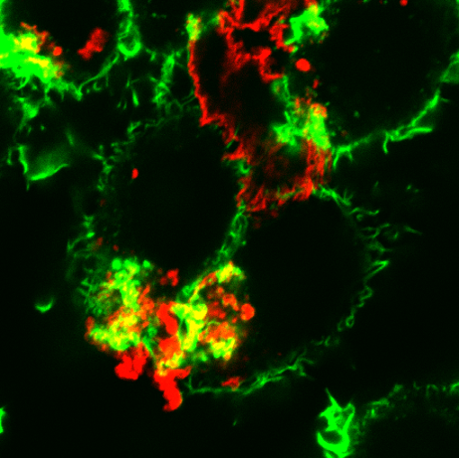
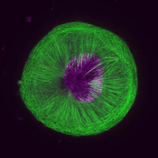
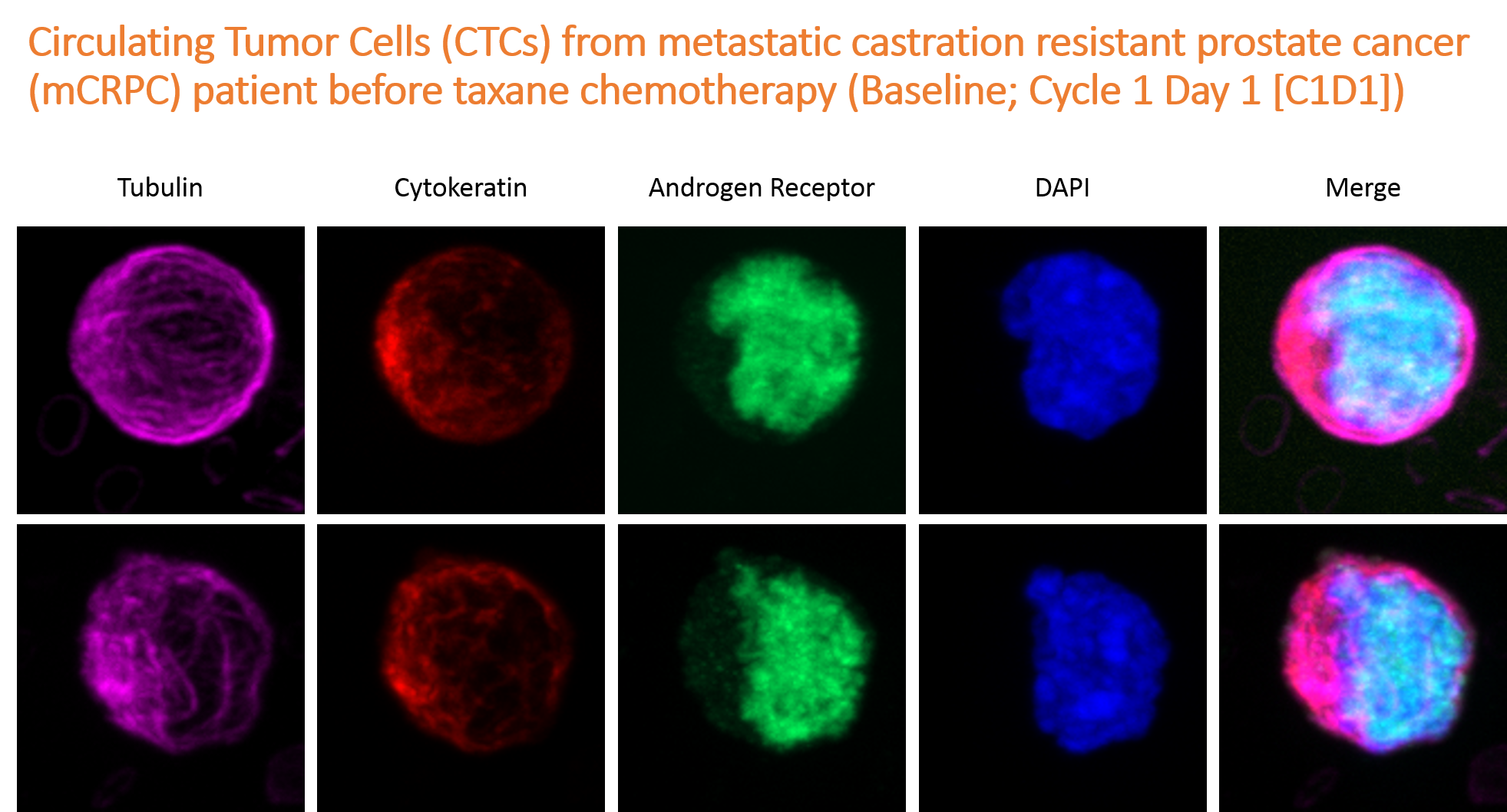
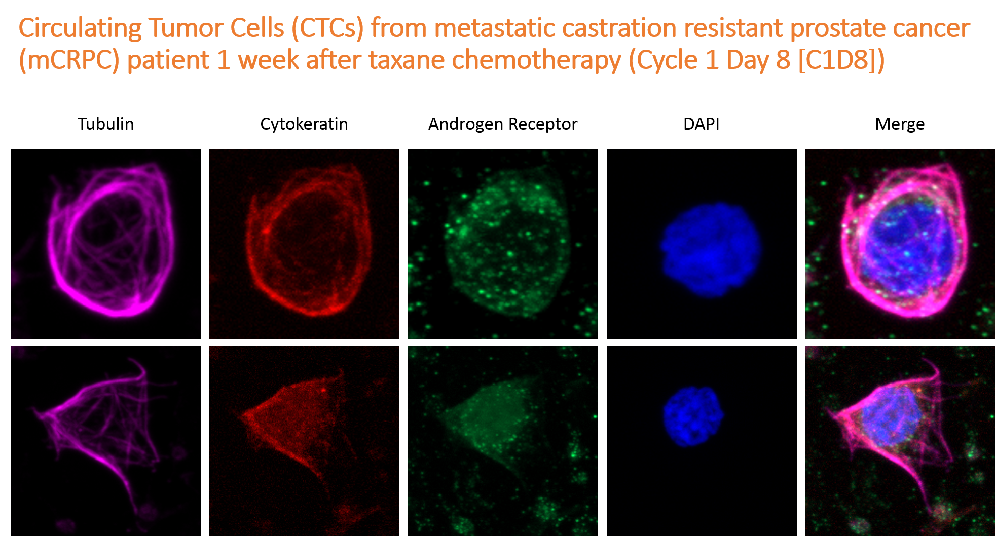
Our Facility offers a variety of microscopes to our users, which are all available for independent, as well as limited assisted use. These include regular widefield microscopes for fluorescence and brightfield imaging, laser scanning microscopes (confocal and multiphoton), super-high resolution microscope (Zeiss Airyscan), automated high content optical microscopy, and electron microscopy.

Image analysis
We provide access to several image analysis software packages, and image analysis workstations that are optimized for handling large amounts of imaging data. The software we have current licence for include MetaMorph, MetaXpress and Imaris. In addition, we have Fiji/ImageJ on all our workstations.

Other services
In addition to microscopy and image analysis, our Core Facility also provides basic histology services. These include sectioning and H&E staining of tissues for light microscopy, and tissue processing for electron microscopy (including sample preparation for Scanning EM and Immuno-EM). In addition, our Core Facility oversees the use of a Phosphorimager and two X-ray film processors.

Education and Consultation
Our Education program consists of training users individually and in small groups. In addition, we offer free consultation of experiment design - including suggesting the best microscope for specific applications, optimal dyes to use, need for specialized imaging chambers etc. Consultation for image analysis are also provided.